Translational Perioperative and Pain Medicine (ISSN: 2330-4871)
ARTICLE DOI: 10.31480/2330-4871/210
Case Report | Volume 12 | Issue 1 Open Access
Anesthesia Management in Endotracheal Laser Surgery: Is it Possible for Complications to Arise Despite Optimal Planning? A Case Report
Denada Haka 1* , Nedim Çekmen 1 , Sinan Issi 2 , Mehmet Dalokay Kiliç 2 and Murathan Ekent 3
1 Department of Anesthesiology and Critical Care Unit, Başkent University Faculty of Medicine, Ankara, Turkey
2 Department of Thoracic Surgery, Başkent University Faculty of Medicine, Ankara, Turkey
3 Department of General Surgery, Başkent University Faculty of Medicine, Ankara, Turkey
Denada Haka, Department of Anesthesiology, Başkent University, Fevzi Cakmak Street. No: 45, Çankaya, Ankara 06490, Turkey, Tel: +90-5054349414Editor: Renyu Liu, MD; PhD; Professor, Department of Anesthesiology and Critical Care, Perelman School of Medicine at the University of Pennsylvania, Center of Penn Global Health Scholar, 336 John Morgan building, 3620 Hamilton Walk, Philadelphia, PA 19104, USA, Fax: 2153495078, E-mail: RenYu.Liu@pennmedicine.upenn.edu
Received: Oct 02, 2025 | Accepted: Dec 20, 2025 | Published: Dec 24, 2025
Citation: Haka D, Çekmen N, Issi S, et al. Anesthesia Management in Endotracheal Laser Surgery: Is it Possible for Complications to Arise Despite Optimal Planning? A Case Report. Transl Perioper Pain Med 2025; 12(2):798-801
Abstract
Endotracheal laser surgery (ELS) is a specialized surgical technique employed in procedures involving the airway with numerous advantages such as microscopic precision, a bloodless operative field, and the preservation of surrounding normal tissue, but also significant risks such as burning, tissue damage, and tracheal rupture. In this case report, we present the case of a 53-year-old male patient undergoing bronchoscopic surgery for laser diathermy treatment due to metastasis of thyroid cancer into the trachea. Considering that both the anesthesiologist and the surgeon share responsibility for the airway, we highlight the critical role of a multidisciplinary approach in managing such cases.
Keywords
Anesthetic management, Endolaryngeal laser surgery, Tracheal surgery, Multidisciplinary team approach
Introduction
Endotracheal laser surgery (ELS) has emerged as a valuable technique in managing airway disorders, including lesions of the larynx and trachea. This minimally invasive procedure offers significant advantages, such as reduced morbidity, shorter recovery times, and the ability to preserve vocal function. However, using lasers in the airway poses risks as they can be invisible, may be misdirected, and could ignite anesthetic gases or endotracheal tubes (ET), potentially damaging healthy tissues [1]. The interaction between the laser and the ET can lead to complications such as fire, airway obstruction, and thermal injury. The all-metal bronchoscope system (Figure 1) utilized for ventilation in ELS offers significant advantages by eliminating the risk of a flammable ET. This system effectively maintains adequate and stable ventilation during the procedure [2]. Consequently, careful planning and execution of anesthetic management are paramount to ensure patient safety and surgical efficacy. In our case, important anesthetic considerations, monitoring techniques, and safety protocols during the ELS procedure are discussed, and the importance of a multidisciplinary team approach in anesthetic management and reducing complications is emphasized. Oral and written consent to publish this case report has been obtained from the patient.
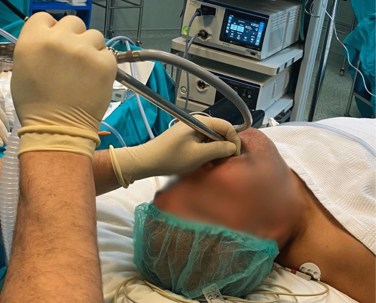
Figure 1: The all-metal tracheoscopesystem.
Case Description
A 53-year-old male patient (weight 82 kg; height 170 cm) was referred to our clinic for the treatment of tracheobronchial lesions utilizing fiberoptic bronchoscopy and laser surgery, followed by a complementary thyroidectomy. His medical history included papillary thyroid carcinoma, for which he had a total thyroidectomy in 2009. Postoperatively, he had remained stable, with no signs of recurrent malignancy, until the recent onset of respiratory symptoms, including hemoptysis and persistent cough over two weeks. A comprehensive evaluation identified a tracheal mass measuring 2.6 cm × 2.7 cm × 3 cm, raising suspicion for possible malignancy. During the preoperative assessment, the patient was classified as an American Society of Anesthesiologists score III due to his previous thyroid cancer and current respiratory symptoms. He exhibited no signs of significant cardiopulmonary compromise, and all laboratory tests were within normal limits. Several anaesthetic considerations were addressed in light of the mass's location and the anticipated use of laser surgery.
The patient's airway was thoroughly evaluated, focusing on potential obstruction from the mass. No dyspnea nor preoperative desaturation were noted. Precautionary measures, including the readiness for difficult emergency intubation and equipment for tracheostomy, were implemented in case of an airway emergency. The use of endotracheal tubes specifically designed for laser surgery was considered. A final decision was to employ the jet ventilation technique with the all-metal tracheoscope instead of apneic ventilation or endotracheal intubation. We acknowledged the potential complications of jet ventilation, including pneumothorax, pneumomediastinum, gastric inflation, resected material aspiration, and mucosal surface dehydration [2]. Preparations for fire risks associated with carbon dioxide (CO2) laser usage were established, including selecting non-flammable materials. All personnel in the operating room were equipped with safety glasses, and the patient's eyes were protected with moist eye pads after being taped. A laser warning sign was placed outside the operating room door to ensure safety for individuals entering the operating room.
Standard monitors were applied when the patient was admitted to the operating room, including an electrocardiogram, pulse oximetry, and noninvasive blood pressure (BP) cuff. ELS was conducted under deep sedation anesthesia. Induction was achieved using a combination of 0.01 mg/kg intravenous (IV) midazolam, 1 mcg/kg IV fentanyl, and a propofol infusion of 3-5 mg/hour IV. The patient's airway was secured with a laser-safe jet mechanical ventilation system with the tracheoscope. Continuous monitoring during the procedure included vital signs, end-tidal CO2, and the oxygen reserve index (ORI) to detect any early desaturation. The surgical team employed a CO2 laser for the precise resection of the tracheal mass. Anesthesia was maintained using total intravenous anesthesia (TIVA) alongside a low dose of muscle relaxants (10 mg rocuronium IV) to minimize straining. The fraction of inspired oxygen (FiO2) was reduced to 21% (to minimize the burning risk), and sevoflurane was administered only before the onset of diathermy, with no volatile agents used during the procedure, relying instead on a mixture of air and oxygen.
During the laser treatment, the tip of the fiberoptic tracheoscope was inadvertently slightly burned, but no additional tissue damage occurred on the patient. Bronchoscopy was subsequently performed to evaluate the extent of any injury, revealing no significant damage. Dexamethasone 8 mg IV was administered to minimize airway swelling. A tracheal stent was placed preoperatively to prevent tracheal stricture (Figure 2). After confirming adequate respiratory function and obtaining normal arterial blood gas values, the patient was transferred to the intensive care unit. He demonstrated stable recovery with no immediate complications and was discharged five days later.
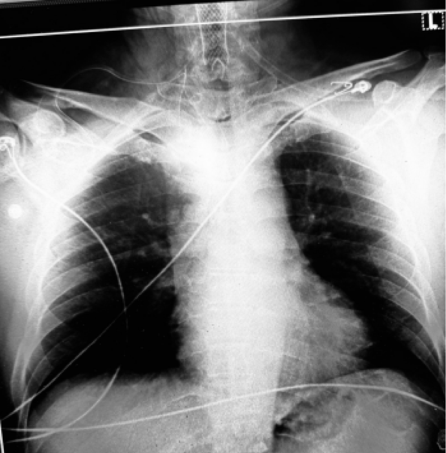
Figure 2: The tracheal stent is seen in chest X-ray.
Discussion
The management of anesthesia in endotracheal laser surgery presents a unique set of challenges, particularly in patients with masses invading the airway. A method of TIVA using short-acting agents was preferred to facilitate early awakening post-surgery and to prevent the aspiration of blood or debris into the lungs [3]. Volatile anesthetics, like isoflurane, sevoflurane, and desflurane, do not support combustion and are not flammable, so that they can be safely used [1]. Ketamine must not be used as it increases airway reflexes like laryngospasm and further obstruction [1]. Applying rocuronium was dangerous for the situated airway but necessary because an ideal anesthetic technique for this surgery requires minimal movement of the vocal cords and a depth of anesthesia sufficient to suppress the hemodynamic response.
Conacher, et al. describe specific anesthetic protocols tailored for laser procedures and prioritized rapid sequence induction intubation [4]. We prioritized laser safety protocols, including a laser-safe, all-metal, non-flammable system. We believe this detail was crucial in preventing the burning at the tip of the flexible bronchoscope from spreading to the tracheal tissue. Conacher, et al. also focused on optimizing ventilation and managing potential complications using arterial line monitorization [4]. We similarly stress the importance of continuous monitoring to promptly detect changes in respiratory status by using ORI. Both studies indicate the significance of vigilant anesthetic management during surgery when it comes to the risk of desaturation and hypoxia.
Divatia J, et al. applied anesthetic management of tracheal surgery using the laryngeal mask airway method (LMA) [5]. The common ventilatory strategies for ELS nowadays include spontaneous breathing, conventional endotracheal intubation, jet ventilation, and intermittent apnea technique. The use of an LMA can minimize airway manipulation during surgery, reducing the risk of complications such as intubation failure, mucosal trauma, bleeding, or perforation, and decreases the likelihood of postoperative complications, including restenosis, anastomotic dehiscence, or dysphonia and lowers the risk of airway irritation and coughing during emergence from anesthesia. However, the tumor in our case was only 1-2 cm below the vocal cords, so our surgeons could not operate with an LMA. On the other hand, plastic ET and LMA are flammable materials and may not be safe for such surgeries.
Even if everything is planned to minimize the risk of urgent explosions, the risk remains. Therefore, preoperatively, a detailed scheme illustrating endotracheal laser surgery safety protocols should be developed. This should include protective gear for healthcare professionals, a depiction of a laser device with clear warning signs, emergency equipment such as fire extinguishers and a first aid kit, proper positioning of surgical instruments, and a checklist of safety protocols (Table 1).
Table 1: Essential of endotracheal laser surgery protocols.
|
CATEGORY |
EQUİPMENT |
PURPOSE |
|
Laser System |
CO₂ Laser, KTP Laser, or Diode Laser |
Precise tissue cutting and coagulation |
|
Endoscopic Equipment |
Rigid or Flexible Endoscope |
Visualization of the airway |
|
Video Camera and Monitor |
Real-time imaging and recording |
|
|
Light Source and Fiberoptic Cable |
Illumination for visualization |
|
|
Endotracheal Tubes |
Laser-Resistant Endotracheal Tube (e.g., Silicone, Metal, or Cuffed with Saline/Methylene Blue) |
Prevents airway fires |
|
Ventilation System |
Jet Ventilation or Low-Flow Anesthesia Circuit |
Maintains oxygenation and reduces fire risk |
|
Fire Safety Equipment |
Wet Sponges, Saline-Soaked Pledgets |
Fire prevention |
|
Suction Catheter |
Removes debris and prevents aspiration |
|
|
CO₂ Fire Extinguisher |
Emergency fire control |
|
|
Surgical Instruments |
Microlaryngeal Instruments (Forceps, Scissors, Suction) |
Assists with tissue handling |
|
Monitoring Equipment |
Pulse Oximeter |
Oxygen saturation monitoring |
|
Capnography |
CO₂ monitoring for ventilation control |
|
Conflict of Interest
None.
Disclosures
Name: Denada Haka MD, Murathan Erkent MD, Sinan Issi, MD
Contribution: This author designed and performed a case report.
Name: Nedim Çekmen and Mehmet Dalokay Kiliç, MD, Professör
Contribution: This author drafted and critically reviewed the article and provided constructive suggestions.
References
- Hermens JM, Bennett M, Hirshman CA (1983) Anesthesia for laser surgery. Anesthesia & Analgesia 62: 218-229.
- Woo P, Vaughan CW (1983) A safe, non-flammable, all-metal, cuffless endotracheal Venturi ventilation system for use in laser surgery. Otolaryngol Head Neck Surg 91: 497-501.
- Scheck PA, Mallios C, Knegt P, Van der Schans EJ (1984) High-frequency ventilation in laser surgery of the larynx. Clin Otolaryngol Allied Sci 9: 203-207.
- Conacher ID, Paes LL, McMahon CC, Morritt GN (1998) Anesthetic management of laser surgery for central airway obstruction: A 12-year case series. J Cardiothorac Vasc Anesth 12:153-156.
- Divatia JV, Sareen R, Upadhye SM, Sharma KS, Shelgaonkar JR (1994) Anesthetic management of tracheal surgery using the laryngeal mask airway. Anaesth Intensive Care 22: 69-73.